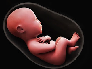
" A BUN IN THE OVEN" - A Journey of Zygote Formation. - Dr. Vandana Patel (PT)
The ovum👩
A single cell released from female reproductive systems, either of the ovaries is called as ovum.
The sperm👨
It is a male reproductive cell which originates from seminiferous tubules of each testicle.
Fertilization👪
All of us are aware about the fusion of male and female gametes which results in the development of a baby in the womb.
Interesting fact is it takes place in the distal third of the oviduct (Fallopian tube)...!!!!
In the above diagram of female gamete all of us can visualise the layer of zona pellucida. Yes, that’s where the fertilization begins. At first when Sperms strikes at the wall of ovum i.e. the zona pellucida, the attachment is quite loose. Eventually, tighter bond is developed with the interaction at receptors located on the surface of zona pellucida. Similarly, sperms have thousand copies of an egg binding protein which can bind with the receptors located at zona pellucida. Following this receptor- sperm interaction, the enzymatic modification takes place within zona which makes sperm to tunnel its way through. The first sperm that penetrates the perivitelline space (between zona pellucida and oocyte plasma membrane) triggers the activation of egg. This activation happens in two steps: first is depolarizing the plasma membrane of oocyte and second is releasing of enzyme from cortical granules which further changes structure of zona pellucida. The sperm that fuses with plasma membrane fertilizes the oocyte and releases its genetic material into oocyte cytoplasm.
The transformation of fertilized oocyte into the three dimensional embryo occurs with well organized cell biologic phenomenon such as cell division, adhesion, secretion, cyto-differentiation, motility and death.
The oocyte undergoes a series of mitotic divisions termed as cleavage. The cells derived from these repeated mitotic divisions are called as blastomeres.
At 8-16 cell stage, blostocyst forms where there is clump of cells with outer layer of trophoblast cells. Once zona pellucida shades off, the blastocyst attaches and implants within uterine cavity. (IMPLANTATION OF EMBRYO) 👶Following this phase; two layers of cell cavity forms. Inner layer called as cytotrophoblast and outer layer syncytial trophoblast which is secretory and critical to implantation.
By second week of gestation, the cells of inner cell mass flatten to form second layer of epithellium. The upper layer locate next to trophoblast is called as epiblast which becomes progenitor of all embryonic tissues and extra embryonic mesoderm and that will represent dorsal side of embryo. These cells will be helpful in forming amniotic cavity. The bottom layer is termed as hypoblast or primary endoderm which forms yolk sac. Thus, two fluid filled cavities develop at the end of second week of gestation without any axial features visible within embryonic disk.
During the third week of gestation, important structure known as primitive streak appears. A midline thickening of epiblast designed to form posterior end of embryo becomes visible. Some epiblast cells enter the streak and get distributed to more ventral regions of embryo as mesoderm and ectoderm. The first cells from streak represent embryonic ectoderm followed by a solid cord of cells, the notochordal process which extend cranially from streak.
The notochord, an axial structure, plays a significant role in formation of nervous system.
Another group of endoderm cells accumulated at one end of primitive streak are termed as the prochordal plate, which is the site of future mouth and thus it marks the cranial region of embryo. Remaining cells forms mesoderm as they lie between ectoderm and endoderm.
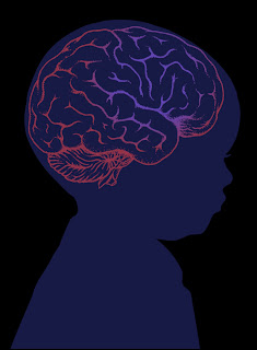
The process of human nervous system development begins at about 18 days after fertilization which makes it first of the organ systems to initiate development.
How is this nervous system being formed in embryonic life..???➽➽➽
Why is it so important for human life..??➽➽➽➽


Cheers. Thank you so much for Sharing this. :)
ReplyDeleteGood content . Waiting for further posts to enrich my knowledge . 💗
ReplyDeleteIt was great 👍, Thanks for sharing the knowledge.
ReplyDeleteAmazing ma'am😍to d point and fab
ReplyDeleteits very informative plus the language is understandable and simple for the non medio commmunity as well.
ReplyDeletesimply perfect.. congratulations for keeping first successful step in blog world...,🌹
ReplyDeleteExcellent writing... Keep up the spirit.... Waiting for more dear👌👌👌👍
ReplyDeleteExcellent work
ReplyDeleteKeep it up vanna
Very rich post. Thanks for sharing.
ReplyDeleteBrowse our website: North Valley Pediatric Therapy