"THE COPPER HEAD" - AN INTRODUCTION TO WILSON'S DISEASE !!! BY DR. VANDANA PATEL (PT)
Wilson’s disease (WD) is an inborn error of copper metabolism that is also known as hepato-lenticular degeneration.
It is an autosomal recessive disorder produced by a mutation on chromosome 13.The gene encodes a transport protein & the mutation of which causes abnormal deposition of copper in the liver, brain (especially the basal ganglia), and the cornea of the eyes.
WD typically begins in childhood, but in some cases has its onset as late as the fifth or sixth decade. About one third of patients present with psychiatric symptoms, one third present with neurological features, and one third present with hepatic disease.
The world wide prevalence of the disease is about 30 in 1 million, with a gene frequency of 1:180.
Normally, the amount of copper in the body is kept constant through excretion of copper from the liver into bile. In WD, the two fundamental defects are reduced biliary transport of copper and an impaired formation of plasma ceruloplasmin. Ceruloplasmin is a plasma ferroxidase. It mediates oxidation of ferrous iron to ferric iron. Due to the mutation extensive changes in copper homeostasis takes place.
The abnormalities in copper metabolism that occur in WD lead to accumulation of the metal in liver and consequently to progressive copper-mediated oxidative hepato-cellular damage; which further leads to liver cirrhosis. Subsequent overflow of copper from the liver produces accumulation in other organs, mainly brain, kidney, and cornea. Within the kidneys, the tubular epithelial cells may degenerate and the cytoplasm may contain copper deposits.
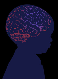
In brain, the basal ganglia show a brick-red pigmentation; spongy degeneration of the putamen frequently leads to the formation of small cavities. There occurs loss of neurons and axonal degeneration. The cortex of the frontal lobe show spongy degeneration. Lesser degenerative changes are seen in the brain stem, the dentate nucleus, the substantia nigra, and the white matter. Copper is also found throughout the cornea.
In the cornea, the metal is deposited in the periphery where it appears in granular clumps close to the endothelial surface of the Descemet membrane. The deposits in this area are responsible for the appearance of the Kayser-Fleischer ring. The color of this ring varies from yellow to green to brown. Copper is deposited in two or more layers, with particle size and distance between layers influencing the ultimate appearance of the ring.
Symptoms and Signs
In most patients, symptoms begin between the age of 11 and 25 years. Onset as early as age 3 and as late as the fifth decade has been recorded.
- Hepatic or hematologic abnormalities
- Behavioral abnormalities
- Neurologic symptoms- Tremor at rest or purposive
- Dysarthria or scanning speech
- Diminished dexterity or mild clumsiness
- Unsteady gait
- Dystonic posturing
- Seizures
- Chorea- small amplitude twitches
- Rigidity and spasms
- Excessive salivation and drooling
- Mental changes
- Episodes of coma
- Keyser- Fleischer ring
Diagnosis:
- Presence of Keyser- Fleisher ring
- Elevated urine copper level- >100µg/24 hrs
- Liver biopsy
Treatment:
- Chelation therapy
- Medical treatments will be directed in the direction of maintenance of copper level in body.
Physical therapy treatment:
Exercises aimed at improving static balance:
Standing exercises on a stable surface, with feet together, semi-tandem and tandem, trying to improve them up to the one-foot standing position. In these positions the patient can be trained with eyes closed trying to modify sensory weighting in favor of somato-sensory information.
Exercises aimed at improving dynamic balance:
on a treadmill under physical therapist’s supervision. Also with wobble board and other dynamic slops.
Exercises aimed at functional capacity training:
Depending upon the activity limitations is the patient the protocol can be decided. For example;
• Walking up and down stairs or ramps
• Picking up objects from the floor
• Unstable surface gait
• Timed up and go test
• 10 minute walk test can be incorporated.
Exercises in severe cases:
• Mild stretching
• Positioning: to overcome attitude of posturing
• Passive movements
• Vestibular stimulations by rocking movements
• Positioning can be advised at home to avoid contractures and muscle shortening.
• If diagnosed in early stage of development treatment to be directed towards achievement of milestones.
Summary:
🤯🤯🤯🤯🤯
References:
Merritt, H. H. (2010). Merritt's neurology. Lippincott Williams & Wilkins.
Daroff, R. B., Jankovic, J., Mazziotta, J. C., & Pomeroy, S. L. (2015). Bradley's neurology in clinical practice e-book. Elsevier Health Sciences.
Maiarú M, Garcete A, Drault ME, Mendelevich A, Módica M and Peralta F. Physical Therapy in Wilson’s Disease: Case Report. Phys Med Rehabil Int. 2016; 3(2): 1082.
Medcomic.com


Your way of explanation is very nice... Keep up the good work !!
ReplyDelete