“THIS LITTLE BUN IS ALMOST DONE” - The Process of fetal brain development – By Dr. Vandana Patel (PT)
“Birth
does not mark a particular milestone in the development of the brain. The brain
continues to develop throughout the lifespan.”
Following fertilization of the ovum,
rapid cell division takes place. As we have already studied during the journey
of zygote formation; that the epiblast cellular layer results in formation of
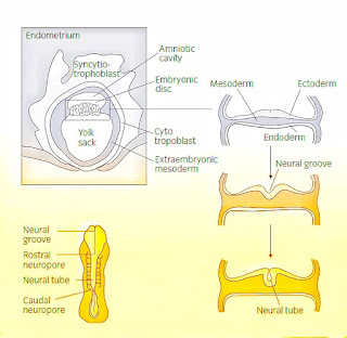
amnionic cavity. From the floor of this amnionic cavity embryonic disc starts
emerging which comprises three layers of cells: the ectoderm, mesoderm and
endoderm.
The inner layer (endoderm) will transform into the internal organs (e.g., digestive and respiratory systems) of the body while the middle layer (mesoderm) will form the musculature and skeletal systems. The outer layer (ectoderm) evolves into a variety of structures of the Central Nervous System (CNS).
The development of CNS begins by thickening of ectoderm layer. This thickening is attributed to increase in the height of ectodermal cells (shape changes from cuboidal to tall columnar) as well as migratory movements of the cells. This thickened layer of ectoderm is known as the neural plate.
Furthermore, two ridges on each side of this neural plate continue to grow, which gives rise to two longitudinal neural folds, forming a neural groove in between.
During this process of neural plate
shaping the broad and short neural plate becomes narrowed transversely and
elongated rostro-caudally. The folds increase in height, curve toward each
other and get fused to form rudiment of the neural tube. This process of formation of initial structure of CNS
is called as neurulation. Neurulation
commences toward the end of the third week of gestation. The neural tube
converts into a cylindrical shape.
After the fusion of neural folds, the formed neural tube separates from ectoderm and becomes buried in the mesenchyme below the surface. During this process, aggregations of cells from the neural folds come to lie on the supero-lateral margins of the tube. They are called as neural crest cells which forms neural crest.
The neural tube grows to form the central nervous
system consisting of the brain and the spinal cord, while the neural crest
forms the peripheral nervous system consisting of autonomic, cranial and spinal
ganglia and nerves. The lumen of neural
tube later becomes the central canal of the spinal cord and the ventricles
of brain.
The head portion of the neural tube becomes the brain while the middle portion becomes the brain stem. The head or cephalic portion further differentiates into the forebrain, midbrain and hindbrain. For instance, by about the fifth week, the neural tube differentiates into the three primary structural units of the brain: the proencephalon (forebrain), the mesencephalon (midbrain) and the rhombencephalon (hindbrain).
By the seventh week two additional structures are formed. The two additional structures are created when the prosencephalon and the rhombencephalon divide in two. The prosencephalon divides into the cerebral hemispheres (the telencephalon) and the thalamus, hypothalamus, epithalamus and pineal bodies (the diencephalon), while the rhombencephalon divides into pons, cerebellum (the metencephalon) and the medulla (the myelencephalon).
Let us conceptualize fetal CNS
development to the construction of a house. The same way that a blueprint
guides house construction, an individual’s genome serves as a blueprint for the
brain. Some of the DNA in the genome creates proteins that build structures,
while others are ‘timing genes’ that manage the sequencing of the building
process. Neurons and glial cells function as the foundational materials of
bricks, wood and cement. Axons, dendrites and synaptic connections among neurons
serve as the wiring for electricity and the telephone.
REFERENCES:
Martin
RP, Dombrowski SC. Prenatal exposures: Psychological and educational
consequences for children. Springer Science & Business Media; 2008 Feb 1.
Polin
RA, Fox WW, Abman SH. Fetal and Neonatal Physiology E-Book. Elsevier Health
Sciences; 2011 Aug 13.
Hepper
P. prenatal development. In Slater A, Lewis M, editors, Introduction to Infant
Development. 2nd edition. Oxford University Press. 2007. p. 41-62






totally like our lecture. classroom recalled ! :)
ReplyDeleteBrilliant writing👌👌... Keep going madam👍👍
ReplyDeleteJzt made as simple as always and amazing 😍
ReplyDeleteSeriously easy and helpful ma'am
ReplyDeleteSeriously easy and helpful ma'am
ReplyDeleteThis was incredibly an exquisite implementation of your ideas, and if you need then come visit us! current issues in the physical therapy field,
ReplyDelete