"LES PETITES MAINS" - DEVELOPMENT OF THOSE LITTLE HANDS PART - I : BY DR. VANDANA PATEL (PT)

Upper
extremity function is important in successfully moving through the activities
of the day. It forms the basis for the fine motor skills important to
activities like feeding, dressing and grooming as well as it plays an important
role in gross motor skills such as crawling, walking, and the ability to
recover balance and protect the body from injury when balance recovery is not
possible. Human hand is useful for prehension, communication and non-
prehensile functions. Effective use of hand depends on the hand skills,
postural mechanisms, visual perception and cognition.
Development
occurs with maturation of different parts of nervous and musculoskeletal
systems as well as with experience.
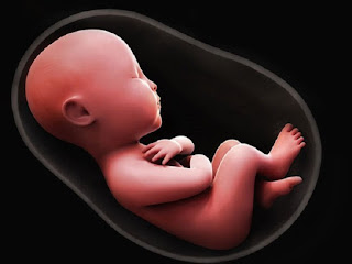
EMBRYOLOGICAL (PRENATAL) DEVELOPMENT
OF UPPER-EXTREMITY:

The
upper limb buds appear on day 24 as small bulges on the lateral body wall at
about the level of C5 to T1. By the end of the 4th week, the upper
limb buds have grown to form pronounced structures protruding from the body
wall. Each limb bud consists of a mesenchymal core of mesoderm covered by an
epithelial cap of the ectoderm. Along the distal margin of the limb bud the
ectoderm thickens to form an apical ectodermal ridge. This structure maintains
outgrowth of the limb bud along the proximal distal axis.
By
33 days the hand plates are visible at the end of the lengthening upper limb
buds. By the end of 6th week, the segments of the upper limb can be
distinguished. Digital rays appear on the hand plates during 6th
week. A process of programmed cell death occurs between the rays to free the
fingers. By the end of 8th week, all the components of upper limbs
are distinct.

The
bones, tendons and other connective tissues of the limbs arise from the lateral
plate mesoderm, but the limb muscles and endothelial cells arise in the somatic
mesoderm and migrate in to limb buds. In general, the muscles that form on the
ventral side of the developing long bones become the flexors and pronators of
the upper limbs. These muscles are innervated by ventral branches of the
ventral primary rami of the spinal nerves. The muscles that form on the dorsal
side of the long bones generally become the extensor and supinator muscles of
the upper limbs. However, some muscles of the limbs shift their position
dramatically during the development.
The
dermatomes of the skin, which represent the tactile system, begin to develop as
early as 7 week of gestation. Proprioceptive system develops in utero with the
differentiation of the articular skeleton and muscle system, beginning around
the 7th week of gestation. The prenatal period ends with full
development of the upper limbs. Incomplete development of the central nervous
system and lack of nerve myelination prevent full upper limb control at birth.
ROLE OF REFLEXES:
The
new born infant is dominated by primitive reflexes that provide most of the
initial response to stimuli from the external world. Tactile and proprioceptive
stimuli elicit the reflexes that most influence early hand function.
These
reflexes are described in table below:

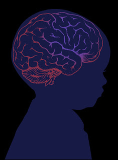
NEURAL CONTROL OF UPPER EXTREMITY:
In primates, neural
pathways controlling movements of the arms are different from those that
control movements of fingers and hand. The two systems develop at different
times.
ARM CONTROL, which is
mainly coordinated at brainstem level,
develops earlier than hand and finger control, which is coordinated at cortical level.

The
capacity to use the hand with skill, in hand– object interactions represents an
evolutionary ability characteristic of the behavior of higher primates.
Three
fundamental prerequisites are necessary for this function:
- Capacity
for independent control over the fingers
- A sophisticated somatosensory system to
guide finger movements
- Ability
to transform sensory information concerning object properties into
appropriate hand configurations.
Each
of these prerequisites is served by separate but interconnected areas of the
cerebral cortex. This includes the primary motor cortex, primary somatosensory
cortex, parietal cortex, and premotor cortex.
Ability to move the fingers individually is thought to result from direct cortico-spinal connections primarily from neurons in the motor cortex to the alpha motor neuron of hand muscles in the ventral horn in the spinal cord.

The
ventral horn of the spinal cord is divided into two main sections, an
inter-neuron zone and the motor neuronal pool or “final common pathway” to the
muscle. The motor neurons in the ventral horn are not randomly distributed but
are clustered into cell columns, a medial cell column that contains the motor
neurons for the trunk, shoulder girdle, and hips, and a lateral cell column
that contains motor neurons for the distal extremities. This direct path is
fast and thought to be important in moving the hand with speed and skill. These
special connections also are thought to be preferentially related to the
intrinsic hand muscles. The intrinsic hand muscles provide the ability to
handle small objects with precision.
Primary
motor cortex is important structure for the execution of independent finger
movements. Damage to motor cortex result in deficit of fine manual coordination.
The
hand is both a motor and sensing organ and there is a tight interplay between
these two functions. The primary somatosensory cortex helps to appreciate
complexity of information processing within this area, particularly for the
hand. Afferent fibres from the dorsal columns project mainly to area 3b for
cutaneous input and area 3a for deep, proprioceptive information. Tactile
information from fingers is necessary to adjust grip to weight and friction of
an object. The transformation of the visual image of an object into an
appropriate hand opening and orientation is processed in the posterior parietal
lobe.
As
the maturation of pyramidal tract occurs at about 9 to 13 months of age,infants
are able to control fractioned finger movements and thus develop more difficult
grasping skills such as pincer grasp.

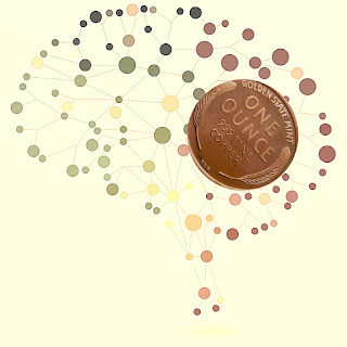
Sensory
inputs from the visual system(eye hand manipulation) go through two parallel
pathways involved in goal directed reaching activities: one related to what is
being reached for (perception and object recognition) and the other related to
where the object is in the space (localisation) and the action system involved
in manipulation of object. The perceptual pathway goes from visual cortex to temporal cortex (ventral
stream pathway), while the localisation and action pathway goes from visual cortex to the parietal lobe (dorsal
stream pathway).
Higher
centres in the cortex take this information and make a plan to generate a
movement. This plan is also sent to cerebellum
and basal ganglia and they modify it to refine movement.
This is just the basics stay tuned for part II to learn more about hand deformities and its management...!!👉👉👉👉
✋✋✋✋✋
References:
Henderson A. & Pehoski C. Hand functions in the child: foundations for remediation. 2nd edition. Elsevier & mosby: 2006; 3-21.
Alexander R., Boehme R. Normal development of functional motor skills; the first year of life. 1st edition; Therapy skill builders: 1993; 11-154.

😍😍best as always
ReplyDeleteNice👌👍
ReplyDeleteSo helpful ...great job..
ReplyDelete